Osteogenesis Type 1
Osteogenesis type I is the most common and usually the mildest form of OI. In most cases, it is characterized by multiple bone fractures usually occurring during childhood through puberty. Fractures usually begin when an affected child begins to walk; fractures during the newborn (neonatal) period are rare. The frequency of fractures usually declines after puberty. Repeated fractures may result in slight malformation of the bones of the arms and legs.
A distinguishing feature associated with OI type I is bluish discoloration of the whites of the eyes (blue sclera). In some cases, individuals with OI type I may develop abnormalities affecting the middle and/or inner ears contributing to, or resulting in, hearing impairment. Hearing loss occurs most often in the third decade of life; however, it can occur as early as the second decade or as late as the seventh.
Individuals with OI type I may have a triangular facial appearance> and an abnormally large head (macrocephaly). Approximately 20 percent of adults with OI type I develop abnormal sideways or front-to-back curvature of the spine (scoliosis or kyphosis).
Additional symptoms associated with OI type I include loose joints, low muscle tone, and thin skin that bruises easily.
Osteogenesis Type 3
OI type II is the most severe type of osteogenesis imperfecta. Affected infants often experience life-threatening complications at, or shortly after birth. Infants with OI type II have low birth weight, abnormally short arms and legs (limbs), and bluish discoloration of the whites of the eyes (blue sclera). In addition, affected infants may have extremely fragile bones and numerous fractures present at birth. The ribs and long bones of the legs of affected infants are often malformed.
Infants with OI type II often have underdeveloped lungs and an abnormally small upper chest (thorax) that may result in life-threatening respiratory insufficiency. In some cases, affected infants may experience congestive heart failure.
Infants with OI type II may also have a small, narrow nose; a small jaw (micrognathia); and an abnormally soft top of the skull (calvaria) with abnormally large soft spots (large fonatanelle). Affected infants may also have abnormally thin, fragile skin and low muscle tone (hypotonia).
OI type II has been subdivided into three subgroups (A, B, and C) based upon small differences in bone formation seen only on x-rays (radiographic features).
Osteogenesis Type 3
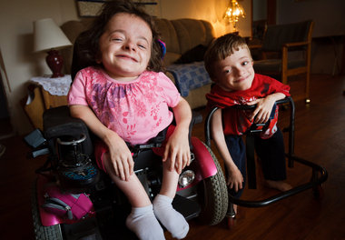
OI type III is characterized by extremely fragile bones, multiple fractures, and malformed bones. Multiple fractures are often present at birth. Fractures and malformation of various bones (most often the ribs and long bones) may become worse (progressive malformation) as affected infants and children age.
Progressive malformation of various bones may result in short stature, sideways and front-to-back curvature of the spine (scoliosis and kyphosis), and malformation of the area where the bone in the back of the skull (occipital bone) and the top of the spine meet (basilar impression). In some cases, affected individuals may develop pulmonary insufficiency and respiratory problems. In severe cases, progressive bone malformation may result in affected individuals requiring wheelchairs.
Infants with OI type III may have a slight blue discoloration to the whites of the eyes (blue sclera) at birth. In most cases, the bluish tinge fades during the first year of life. Affected infants may have a triangular facial appearance due to an abnormally prominent forehead (frontal bossing) and an abnormally small jaw (micrognathia). In some cases, hearing impairment and brittle, discolored teeth (dentinogenesis imperfecta) may also be present.
Osteogenesis Type 4
Individuals with OI type IV have fragile bones that often fracture easily. Fractures are more common before puberty. Affected individuals experience mild to moderate bone malformation and are usually shorter than average. Affected individuals may develop sideways and front-to-back curvature of the spine (scoliosis and kyphosis).
Individuals with OI type IV may have a triangular facial appearance. In most cases, the whites of the eyes (sclera) are normal or pale blue during infancy. As an affected infant ages, the pale blue discoloration of the sclera fades. Affected individuals may also experience hearing impairment and brittle, discolored teeth (dentinogenesis imperfecta)
Unclassified Osteogenesis Imperfecta
Many cases of individuals with the bone abnormalities characteristic of osteogenesis imperfecta have been reported in the medical literature. However, these cases have additional symptoms that prevent them from being classified under one of the four main types of OI. Researchers have speculated that these cases may be subgroups of one of the four main types of OI, an additional type of OI (e.g., OI type V), or separate disorders.
Your doctor can diagnose brittle bone disease by taking X-rays. X-rays allow your doctor to see current and past broken bones. They also make it easier to view defects in the bones. Lab tests may be used to analyze the structure of your child’s collagen. In some cases, your doctor may want to do a skin punch biopsy. During this biopsy, the doctor will use a sharp, hollow tube to remove a small sample of your tissue.
Genetic testing can be done to trace the source of any defective genes.